Horse Hoof Anatomy, Part 1 - Christy West, www.thehorse.com, Dec 2019
The angle between the bottom (solar) surface of the coffin bone and the front of the bone is usually around 50 degrees, but it can be steeper (usually in a club foot) or flatter (usually in a long-toe, low-heeled foot). The coffin bone can fracture and remodel significantly in response to stress. Loss of the distal rim (toe edge) of the coffin bone can occur if there isn’t much foot or sole mass (for shock absorption and protection); this can be secondary to (caused by) poor foot growth, lack of sole thickness/protection, or club foot.
"The palmar angle, between the bottom or wings of the coffin bone and the ground, can be a significant indicator of foot health. For example, a normal palmar angle is slightly positive (heels slightly higher than the toe). But Rucker says an angle higher than 10° suggests the club foot is severe or laminitis has caused instability of the bone within the hoof (and severe laminitis can cause coffin bone breakdown starting at the rim and even significant loss of bone mass). Conversely, a zero or negative palmar angle (heels lower than the toe) suggests that the foot has crushed heels and a compromised digital cushion."
The collateral (or lateral) cartilages extend upward from either side of the coffin bone and help the hoof expand with weight bearing. Ossification of the collateral cartilages can limit the hoof’s natural expansion and result in some sensitivity to touch via hoof testers. Lameness isn't always seen, and this is most commonly seen in heavy horses working on hard ground.
The digital cushion lies below the coffin bone and above the frog and cushions the foot from ground forces. When a horse has a broken back hoof pastern angle and/or long toe/low heels and the heels are over-loaded, the digital cushion is crushed. Stephen O'Grady says "The most common problem I see in internal hoof structures is poor heel structure and inadequate sole depth (due to digital cushion loss)”.
Between the coffin bone and the hoof wall lies the corium - soft tissue that includes blood vessels, nerves, and the laminae—interlocking leaflike structures that attach the hoof to the bone. If a disease or trauma causes swelling in the hoof, the hoof can’t expand to accommodate both the swelling and the blood flow, and swelling generally wins. Without blood flow, tissue and bone can die. Laminitis is disease of the laminae that can range from mild to severe and transient to chronic, often caused by metabolic problems like Equine Metabolic Syndrome and PPID (in both cases insulin dysregulation leads to laminitis). The laminae can detach and the coffin bone rotate away from and sink down within the hoof capsule.
Hoof Trimming to Improve Structure and Function - notes from Robert Bowker 2019 NEAEP Symposium, Stephanie Church, www.thehorse.com, Dec 2019
Dr Robert Bowker, longtime podiatry researcher and former professor and head of the Equine Foot Laboratory at Michigan State University’s (MSU) College of Veterinary Medicine, described his perspectives and trimming approaches during a presentation at the Septenber 2019 NEAEP symposium.
The foot should be balanced approximately 50:50 toe:heel, so that if a perpendicular line is dropped from the center of rotation of the short pastern bone (P2), half the foot is in front of the line and half behind.
When a horse with peripheral thinning of P3 gets laminitis and rotation, the bone cannot support the weight of the horse and becomes crushed.
Bowker trims to shorten the toe, trimming inside the white line if necessary, and promote caudal (toward the rear) migration of the heels to bring the central sulcus (the cleft between the heels) back to the sole of the foot so it makes light contact with the ground. He said trimming with these goals can improve the foot’s health and get the ratio to approach 40:60—allowing the back part of the foot to enlarge and return to its robust health.
To correct a long toe/underrun heels foot:
Bring the heels back to the level of (back of) the frog, and so that the frog just kisses the ground - too much pressure reduces the blood supply and causes the frog to atrophy.
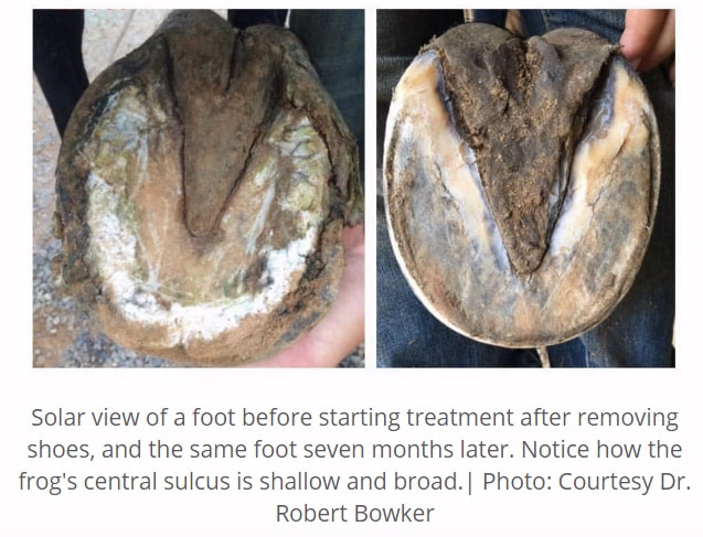
The frog's central sulcus should be wide and shallow, and ideally the frog should not be trimmed, as trimming causes the frog to retract and reducees its ability to dissipate energy [however the frog may need to be trimmed if heels need to be lowered].
Bevel the toe from beneath (the sole), not the outside (dorsal hoof wall), trimming inside the white line every few days to keep the toe short.
Trim the foot initially every 3-4 days until the toes and heels are back under the horse, then the trim interval can be lengthened to up to 4 weeks (less when feet are growing quickly).
Measure the feet every 3-4 days to monitor changes.
Bowker has been able to improve digital cushions despite wide acceptance that digital cushion damage is permanent (it isn't) - he says "internal changes can occur if the farrier or trimmer gives the foot an opportunity. A crushed digital cushion will repair itself with myxoid cells", and "you can always improve the trim to improve the internal structure of the tissues. If you have a short toe, you'll have a pretty good foot".
"Here are some insights and tips Bowker shared on the normal equine foot and what goes wrong with typical husbandry practices:
1. The foot is extremely adaptable. Conformation is a point in time. This can be corrected and improved if the foot is given the opportunity.
2. The foot adapts to the environment (trimming, shoeing, loading, ground surface, moisture, etc.), but the biggest environmental factors are the hoof care professional and the trim. “They can cause major environmental effects and greater biomechanical changes in a brief time period. These changes affect the internal tissues—they respond each time the foot is trimmed.”
3. All feet are different, even in the same horse and in the same pair of legs.
4. Long toes and underrun heels are ubiquitous, and much of the horse industry accepts them as normal. “I have several thousand sagittal photos of feet … (only) one of them is (balanced) 50:50 front to back,” he said.
5. As the toe and coffin bone lengthen, Bowker says the coffin bone remodels internally as it attempts to support the longer toe; the bone becomes more porous due to more movement between the bone and hoof wall. “This increased porosity is not beneficial”.
6. Navicular bone movement up and down, due to the changes in foot mechanics, damages the deep digital flexor tendon, as well as insertion ligaments of the distal sesamoidean impar ligament.
7. With long-toe, underrun heels, the bottom end of the short pastern (P2) changes shape, too. It starts out symmetrical in its articulation with the coffin bone. But current standard trimming methods alter the biomechanics, as the ends of P2 become asymmetrical. “With the gradually increasing length of the coffin bone, chip fractures begin to appear on the navicular bone, and they can appear (at any age). Many associate these fractures with navicular syndrome. If you have a short toe, you don’t have that.”"


 RSS Feed