Feet A-Z
Apex (true apex of frog)
Bars
Bevel/mustang roll
Blood supply/circulation
Bowker, Robert
Breakover
Bruising
Bullnose
Collateral grooves
Coronary band
Cracks
Daisy Haven Farm/Daisy Bicking
Frog
Healthy feet
Heels
Hoof boots
Hoof growth rate
Hoof Pastern Axis (HPA)
Horn:laminar zone
Laminar/lamellar wedge
Mustang roll - see Bevel
Navicular
Palmar/plantar angle
Ramey, Pete
Rings and ridges
Shoes
Ski tip
Sole
Taylor, Debra
Thrush
Toe
Walls & wall flare
White line/White Line Disease (WLD)
Widest part of the foot
|
Apex (true apex of frog)
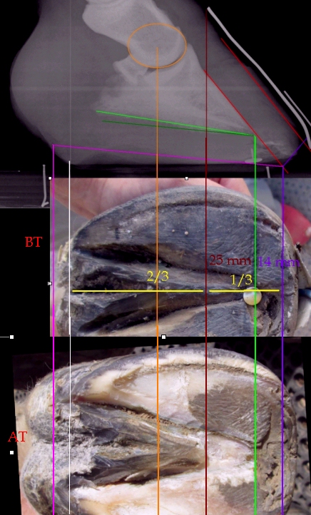
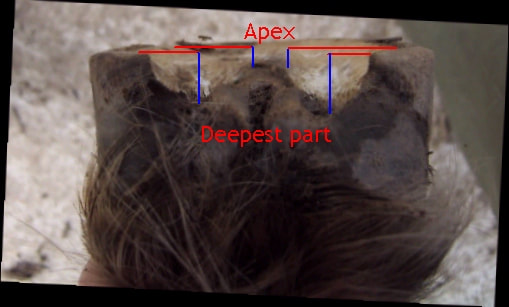
The true apex of the frog should be found by carefully trimming the frog until the line of dirt between the frog and the sole begins to disappear and the "seam" between the sole and the frog is seen. In Page, Bowker, Ovenik & Hagen AAEP 1999 How to Mark the Foot for Radiography the authors find the true frog apex "by trimming the superficial tissue at the apex of the frog to observe the frog blending to the sole. This transitional zone is marked by a color difference; the frog is darker, the sole lighter." Fig 1 in the paper illustrates this colour difference. Following laminitis, the frog often stretches forward. A line dropped down from the hairline at the toe will usually indicate the approximate position of the true apex of the frog. What is thought to be the apex of the frog should be marked when radiographs are taken so that the true and actual frog apex can be compared. In the diagram on the right, four months after laminitis and rotation the frog had stretched forwards. Radiographs were taken with the actual frog apex marked with a drawing pin, and the hairline at the toe marked. On the radiograph the actual apex was clearly too far forwards - the true apex of the frog is always behind the tip of P3. On the radiograph a line was dropped down from the hairline at the toe (brown line) to indicate the approximate position of the true apex. A photograph of the sole taken at the time of the radiograph was adjusted to the same scale as the radiograph, and the brown line suggesting the position of the true apex projected onto the photo of the sole. This helped to guide the farrier's exploration of the true apex. By gently scraping back frog material, the farrier found the clear seam between the sole and the frog - the true apex - as shown in the bottom photo of the sole (brown line). When correctly trimmed, approximately 2/3 of the foot will be behind the true apex, and 1/3 in front of it. The frog stretching forwards like this is often the reason that horses' toes are left too long. |
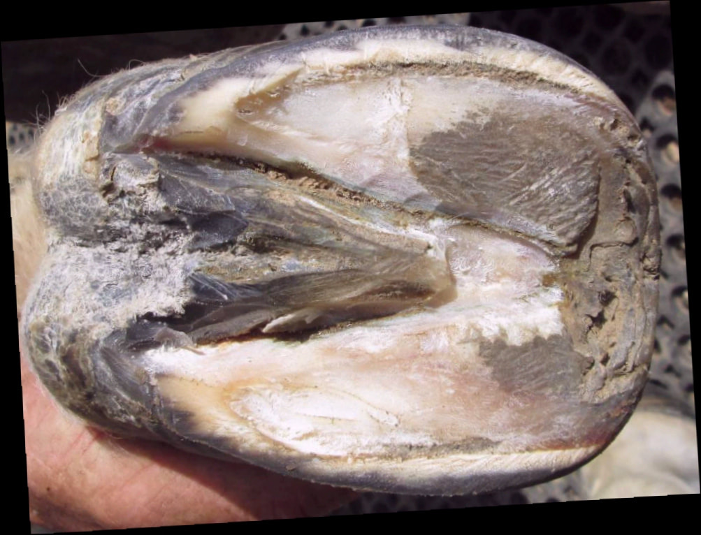
The transition from sole to frog is marked by a colour difference - the frog is darker.
|
|
Bars
Inside a Laboratory that Looks Inside Horses’ Hooves - Dr Lisa Lancaster Robert Bowker has suggested that the bars have tertiary as well as primary and secondary epidermal laminae, and that the bars' laminae "appear able to form tubular horn and contribute cells to the growth of bone the hoof wall and the sole". "Recognizing deformity or malfunction in the bars may turn out to be an important first step in restoring symmetry and function." The Bars - Pete Ramey |
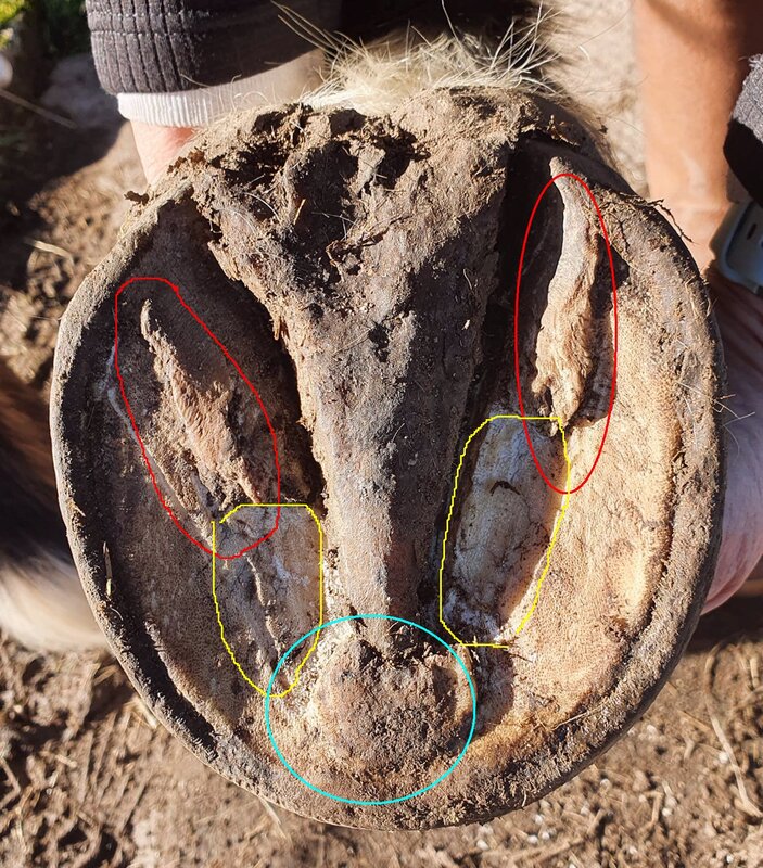
Following damage and sensitivity to the frog, the bars grew longer on this foot, seeming to surround the frog. Red = bars, yellow = bar material that has popped off, turquoise = ridge of harder bar (?) material not yet ready to pop off.
|
Bowker, Robert
Long Toe Woes: Perspectives on Navicular with Dr. Robert Bowker - The Humble Hoof, May 2020
Hoof Trimming to Improve Structure and Function - notes from Robert Bowker 2019 NEAEP Symposium, Stephanie Church, www.thehorse.com, Dec 2019
See also: Articles about feet December 2019
Physiological Trimming for a Healthy Equine Foot by Dr. Robert Bowker
Equine Foot Laboratory - Michigan State University
Robert Bowker Hoof Anatomy videos 1-7
Breakover
Breakover is defined as the most dorsal location of the solar aspect of the hoof capsule that contacts the ground, and the last part of the hoof to leave the ground as the horse moves.
In Page, Bowker, Ovenik & Hagen AAEP 1999 How to Mark the Foot for Radiography, 30 lame horses were shod or trimmed on the front feet with breakover 4 mm (for horses weighing 200-300 kg), 5 mm (300-400 kg), 6 mm (400-500 kg) or 7 mm (500-600 kg) in front of a line dropped down perpendicularly from the tip of the pedal bone (P3) shown on radiographs, with this distance measured on the radiographs from a pin at the true frog apex and plotted on the foot. On the hind feet breakover was set at the above distances plus 1 mm. For barefoot horses, the toe was rolled at a 15 degree angle finishing at the breakover line on the foot. This trim protocol brought breakover back by an average of 15 mm (mean 38 mm from pin to breakover before treatment, 23 mm from pin to breakover after treatment). 15/30 horses showed an improvement of 1 lameness grade within 1 hour, and 22/30 had improved 1 grade after 6 weeks. Bringing breakover back caused improvements in the hoof pastern axis/angle between P1 and P3. Bringing breakover closer to the tip of P3 decreased the length of the lever between breakover and the DDFT insersertion, decreasing the strain on the DDFT. Beware bringing breakover TOO far back as that may make the hoof pastern axis too high.
Jenny Edwards of All Natural Horse Care suggests that breakover should be in the position that puts the least stress on the laminae, and therefore the point of breakover should start at the white line - Toe Rocker - all-natural-horse-care.com
The widest part of the foot - Gene Ovnicek
Gene Ovnicek suggests that breakover should be around 1/4" in front of the tip of P3.
Page BT, Hagen TL
Breakover of the hoof and its effect on structures and forces within the foot
Journal of Equine Veterinary Science June 2002 Vol 22 Issue 6 pages 258-264 (Full paper)
"The location of the breakover of the hoof capsule can be positioned through shoe placement, shoe shape, or trimming on barefoot horses. Placing the breakover in relation to the tip of PIII is a more dependable location than placing breakover guided only by visual evaluation of the hoof capsule. The hoof-pastern axis and the position of the navicular bone will be affected by the distance from the apex of the frog to breakover. The resulting decrease in strain of the deep digital flexor tendon while standing and during movement [presumably from bringing breakover back] will decrease inflammation and disease in the equine digit."
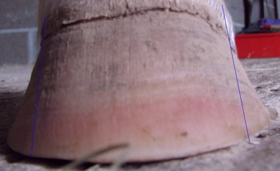 Bruising in the hoof wall - this pony had had laminitis, but also had a change of angle in the hoof wall and flaring.
Bruising
See also White Line - Bruising/blood in the white line - below.
Red colouration indicating bruising may be seen on white hoof walls and soles, or in the white line.
When small blood vessels break in the sensitive tissues of the foot, blood seeps out into the surrounding tissue, and if trapped between the hoof wall or sole and the tissue, it cannot be absorbed and will grow down with the hoof wall/sole, eventually being seen as a bruise. A sole bruise may be seen approx. 4 to 8 weeks after the injury occurred. A bruise on the hoof wall or in the white line may be due to injury to the laminae or coronary band, and may be seen up to 9 months after the injury occurred.
Bruises from coronary band injury may be hidden by the periople initially so may not be seen until several weeks after the injury took place.
Bruising may be caused by laminitis, but also by poor foot balance or by trauma/injury to the foot or coronet.
How to prevent stone bruises - Susan Kauffmann Sept 2018
Hoof bruises - Paige Poss - Iron Free Hoof
Tending to a tender foot - Karen Briggs www.thehorse.com Oct 2001
Bullnose
Barefoot Horse Blog - Laminitis - what is going on inside? Good example of exterior of hoof showing bullnose and the same hoof dissected to show rotation and laminar wedge.
Barefoot Horse Blog - Bull nosed hooves
According to Marjorie Smith, a bullnose is more often seen on a hind foot which has a long toe and short heel - the mechanical forces on the hoof pull the wall away from the bone, stretching the white line - www.barefoothorse.com - Flares.
White Line Tightens Up After Shoes Are Removed - Christina - barefoothoofcare.net 2013
Collateral grooves
The collateral grooves (CGs) are the grooves between the sides of the frog and the sole. Measuring the collateral grooves gives a guide to the thickness of the sole and the height of the pedal/coffin bone off the ground, as the collateral grooves are a reasonably consistent distance from the corium of the pedal/coffin bone. The pedal/coffin bone is covered by a 2 to 6 mm "sponge" of blood vessels and nerves called the corium; the sole protects the pedal/coffin bone and the corium from the ground, and should be at least 10 mm thick at the edge of the sole (next to the white line). According to Pete Ramey, "the bottom of the collateral groove is generally 8 mm away from the corium" (reference: Understanding the horse's sole - Pete Ramey, also Care and Rehabilitation of the Equine Foot - Pete Ramey), so around 10 to 14 mm away from the pedal/coffin bone.
See Measuring Collateral Grooves
The collateral grooves are measured towards the back of the frog where the groove is deepest, and at the true frog apex (but be aware the frog often runs forward following laminitis)
|
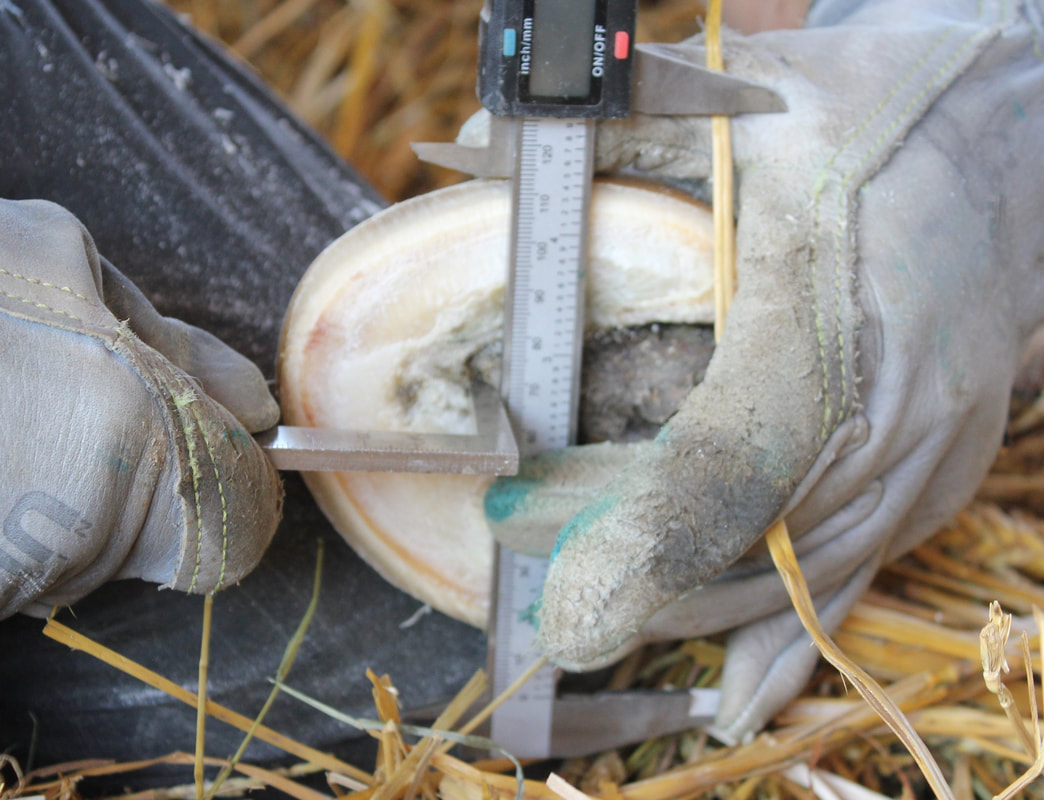
Place a flat object across the foot, and measure from the bottom of the collateral groove to the bottom of the flat object (taking off any wall height above the edge of the sole).
|
Coronary Band
Eustace RA, Emery SL, Cripps PJ
A Retrospective Study of 23 Cases of Coronary Band Separation Longer than 8 cm as a Sequel to Severe Laminitis
Journal of Equine Veterinary Science April 2012 Vol 32 Issue 4 pages 235-244. https://doi.org/10.1016/j.jevs.2011.08.021
Eustace RA, Emery SL, Cripps PJ
A Retrospective Study of 23 Cases of Coronary Band Separation Longer than 8 cm as a Sequel to Severe Laminitis
Journal of Equine Veterinary Science April 2012 Vol 32 Issue 4 pages 235-244. https://doi.org/10.1016/j.jevs.2011.08.021
Daisy Haven Farm/Daisy Bicking
Useful/interesting articles:
How To Develop A Healthy Foot: Circulation Is It - Daisy Bicking
When Your Frog is Down: Repairing Prolapsed Frogs - Daisy Bicking 2014
Useful/interesting videos:
Using the Rasp
Applying Glue-on Composite Shoes for a Laminitic Mare (video)
Artimud video: Foot prep
Useful/interesting articles:
How To Develop A Healthy Foot: Circulation Is It - Daisy Bicking
When Your Frog is Down: Repairing Prolapsed Frogs - Daisy Bicking 2014
Useful/interesting videos:
Using the Rasp
Applying Glue-on Composite Shoes for a Laminitic Mare (video)
Artimud video: Foot prep
Hoof Mapping: Daisy Haven Farm Style - posted on YouTube by Daisy Haven Farm 18 December 2019
Laminitic/Foundered Hoof Trim: Retained Bar & Welded Frog - posted on YouTube by Daisy Haven Farm 17 April 2022.
Exfoliating excess frog and bar/sole material around the frog on a chronic laminitic hind foot.
Exfoliating excess frog and bar/sole material around the frog on a chronic laminitic hind foot.
Horse with Long Hooves Transitions to Barefoot, Hoof Mapping, Trimming Behind the White Line - posted by Daisy Haven Farm on YouTube 07 February 2023
Hoof Mapping Example: Chronic Laminitis with LONNNNNG Frog - posted by Daisy Haven Farm on YouTube 25 November 2023
Sure Foot webinar No. 192 Daisy Bicking live from the barn trimming a laminitis horse - posted on YouTube by SURE FOOT Equine Stability Program 15 April 2021
Sure Foot webinar No. 181 Daisy Bicking Preventing Laminitis, A Farrier's Perspective - posted on YouTube by SURE FOOT Equine Stability Program 23 March 2021
Sure Foot webinar No. 155 Daisy Bicking Sole Mapping - posted on YouTube by SURE FOOT Equine Stability Program 02 February 2021
Sure Foot webinar No. 119 Daisy Bicking talks about the hoof's influence on Balance, Posture and using glue-on shoes - posted on YouTube by SURE FOOT Equine Stability Program 28 October 2020
Sure Foot webinar No. 11 Daisy Bicking - posted on YouTube by SURE FOOT Equine Stability Program 14 April 2020
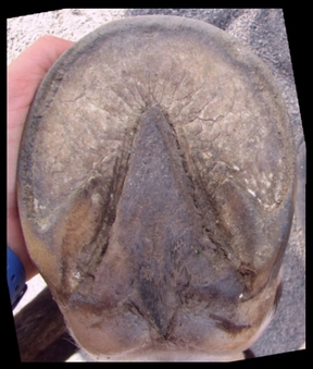
Frog
Healthy frogs make healthy bare feet - Linda Cowles www.healthyhoof.com
"Most of the time the width of the true sole in front of the frog will be about 1/3 of the overall length of the frog" - p 344 Care and Rehabilitation of the Equine Foot - Pete Ramey.
Prolapsed Frogs
When Your Frog is Down: Repairing Prolapsed Frogs - Daisy Bicking 2021
Daisy defines a prolapsed frog as being where the frog, frog corium and digital cushion have fallen below the heels - it is a displacement of the soft tissues in the back of the foot, a collapse of the heel arch.
A horse with a prolapsed frog may be lame, land toe first, be sensitive to pressure on the frog, have a weak digital cushion, have weak, laid over bars and folded over heels, have a low toe wall angle, a bulge in the heel quarters, a broken back hoof pastern axis, a negative palmar angle and suffer from heel abscesses.
Working to radiographs, Daisy aims to rebalance the foot and re-establish a 3 to 8 degree palmar angle, 50:50 base of support either side of the centre of rotation, hoof capsule alignment to the pedal/coffin bone, a straight hoof pastern axis, and to reduce flare and distortion, and she builds a prosthetic heel e.g. with a composite shoe and dental impression material to give caudal support while the hoof corrects. Dental impression material can also be used inside a boot. Any frog infection should be treated, e.g. with CleanTrax.
Heels
A healthy foot will have relatively low heels.
Heel Height: The Deciding Factor - Pete Ramey
Raising the heel/increasing heel height
An article with dissections explaining how high heels increases pressure on the tip of P3:
Uniform Sole Thickness - MT Savoldi, GF Rosenberg
Equine Veterinary Journal, 43: 536–542 2011
The effect of hoof angle variations on dorsal lamellar load in the equine hoof
Ramsey G, Hunter P, Nash M
Reasons for performing study: In the treatment of laminitis it is believed that reducing tension in the deep digital flexor tendon by raising the palmar angle of the hoof can reduce the load on the dorsal lamellae, allowing them to heal or prevent further damage.
Conclusions and potential relevance: The models in this study predict that raising the palmar angle increases the load on the dorsal laminar junction. Therefore, hoof care interventions that raise the palmar angle in order to reduce the dorsal lamellae load may not achieve this outcome.
Vet Clin North Am Equine Pract. 2010 Aug;26(2):391-405. (PubMed)
Clinical presentation, diagnosis, and prognosis of chronic laminitis in Europe.
Eustace RA.
Raising the heels will put the alignment of the phalangeal bones further out of alignment, risks increasing compression at the coronet and further shearing of the laminae at the toe, leading to further rotation.
Heel first landing
Debra Taylor and Pete & Ivy Ramey advocate returning a horse to turnout and in-hand exercise following laminitis when they "have been trimmed, stabilized and have a heel first landing in boots" (Hoof Rehabilitation Protocol - Debra Taylor, Pete & Ivy Ramey). So it's important to recognise a heel first landing:
The anatomy of heel first and toe first landing - Nic Barker, Rockley Farm, Oct 2011
The horse, the "experts" and the heel first landing - Nic Barker, Rockley Farm, Aug 2013
Lots of examples, including video, of horses landing heel first, and why it is so important.
Why Does My Horse Land Toe-First? Pete Ramey, Horseback Magazine, 2013
This video from Alicia Harlov of The Humble Hoof explains how to assess whether a horse is landing heel first or toe first:
Below - Heel First Landings with The Horse PT Dr Barbara Parks:
Slippering heels
DHF Addressing Hoof Distortion: Slippering Heels
Underrun heels
In this short video Bernd Jung of HUFCHECK explains with a simple animation what underrun heels are:
DHF Addressing Hoof Distortion: Slippering Heels
Underrun heels
In this short video Bernd Jung of HUFCHECK explains with a simple animation what underrun heels are:
What are underrun heels ? from HUFCHECK media on Vimeo.
Hoof boots
Why use hoof boots?
Boots and Pads: A True Breakthrough In Healing - Pete Ramey
See "Foundered Horses: Support, Stimulation and Pain Relief" towards the end of the article.
Hoof boots with pads were the first choice for sole protection for the 14/14 obese horses with laminitis returned to their pre-laminitis level of soundness:
Taylor D, Sperandeo A, Schumacher J, Passler T, Wooldridge A, Bell R, Cooner A, Guidry L, Matz-Creel H, Ramey I, Ramey P
Clinical Outcome of 14 Obese, Laminitic Horses Managed with the Same Rehabilitation Protocol
JEVS published online 05 Feb 2014
Hoof Boots - hoof protection for shoeless horses - www.all-natural-horse-care.com
Hoof Boots Overview - www.healthyhoof.com
Where to buy hoof boots:
UK
Equine Podiatry Supplies - new boots
The Saddlery Shop - new boots
Hoof Bootique - new and secondhand boots and boot hire
Urban Horse - new boots and hoof care/trimming equipment
Cannock Chase Equine - new boots
Atlantic Equine
See also TLS forum: hoofboots - secondhand and new
Makes of hoof boots:
Cavallo
EasyCare Inc - Easyboots
Equine Fusion
The Tubbease Hoof Sock may be useful for soaking feet e.g. for abscess treatment and for short-term protection.
Boots that come in small sizes:
Equine Fusion boots start from 66-75 mm in length, and can be used for riding and turnout - see The Saddlery Shop
The Easyboot mini range includes 4 sizes which start from 44 mm wide x 49 mm long and increase to 76 mm wide x 86 mm long - see Easyboot Mini.
The Cavallo Cute Little Boots include 4 sizes which start from 51 wide x 62 mm long and increase to 92 mm wide x 102 mm long - see Cavallo Cute Little Boots.
Easyboot Transition boots start from 89 mm wide x 93 mm long and can be used for turnout/rehabilitation and light hacking - see EasyCare Inc
www.naturalhorsetrim.com boot swap page:
Descriptions and sizing for most makes of hoof boots, and boot swap adverts at the end of the page
Hoof growth rate
Hoof growth in normal & laminitis horses - Tom Ryan FWCF
In mature horses, hoof wall grows from the coronary band at an average rate of 8 - 10 mm per month. This rate can be affected by different factors, including breed, age, nutrition and environment.
Lewis C, Nadeau J, Hoagland T, Darre M
Effect of Season on Travel Patterns and Hoof Growth of Domestic Horses
Journal of Equine Veterinary Science Volume 34, Issue 7, July 2014, Pages 918–922
Hoof Pastern Axis (HPA)
The hoof pastern axis compares the angle of the pastern bones to the angle of the toe/pedal bone (the toe should always be parallel to the dorsal surface of the pedal bone). There should be a straight line through the bones.
The Truth about Hoof Pastern Axis - The Equine documentalist April 2020
Broken back/broken forward HPA
Navicular syndrome - www.horsevets.co.uk - broken back HPA marked on x-ray
Proper Physiological Horseshoeing: What is it? How Do We Apply It? - Stephen E O'Grady - AAEP 2009 - article shows a parallel HPA, a broken back HPA, and describes broken back and broken forward HPAs (inclusion of this article should not be taken as endorsement of the shoeing methods described).
Broken Back Hoof/Pastern Axis - www.thenaturalhoof.co.uk
Hoof pastern angles - Rockley Farm
Horn:laminar zone
The distance between the dorsal surface of P3 and the outer surface of the dorsal hoof wall is called the H:L zone or horn:laminar zone or wall thickness. The H:L zone may widen with conditions that affect the laminae and wall thickness, such as laminitis (the lamellar zone widens) and white line disease (the horn zone widens).
The H:L zone can only be measured accurately if the inside (medial quarter) of the foot was in contact with the x-ray cassette when the lateral (LM) x-ray was taken so that there is no magnification, or if the image has been corrected for magnification. "The thickness of the H:L zone will be randomly and unpredictably magnified when the medial quarter is not touching the cassette during image acquisition" (p 240 Care and Rehabilitation of the Equine Foot 2011 - ch. 13 Radiographic Imaging of the Laminitis Patient - Dr Debra Taylor). The dorsal hoof wall must be accurately marked with radiopaque material such as barium for H:L zone measurements to be accurate.
The H:L zone is measured with the ruler at 90 degrees to the dorsal surface of P3, and can be measured anywhere along the dorsal surface of P3, but usually proximal (just below the extensor process) and distal (just above the tip of P3) measurements are taken and recorded e.g. 18/18 (proximal/distal). In an adult foot with no rotation, the proximal and distal measurements should be the same. In a foot with rotation, the distal (lower) measurement is likely to be greater than the proximal (upper) measurement.
M.C.F= Actual length of marker / radiographic length of marker so the true distance = length measured radiographically x M.C.F
Linford et al 1993 suggested that wall thickness in the normal horse is less than 30% (25 - 30%) of the length of the palmarcortical (PCL) of P3.
"Normal" H:L zones for different breeds have been suggested (using a true lateral radiographic image with no magnification):
Light breeds e.g. Quarter Horse, Thoroughbred 15-16 mm (Clinical and Radiographic Examination of the Equine Foot, RF Redden, AAEP 2003).
Standardbred 20 mm (Clinical and Radiographic Examination of the Equine Foot, RF Redden, AAEP 2003).
Thoroughbred 16.3 mm (Cripps and Eustace 1999).
Dareh-Shori horses (Iran) 17.1 mm (Vali R 2014).
The H:L zone measurement will depend on the size of the foot - "an appreciation of the range of normal for that type and size of horse is essential for accurately interpreting this area" (Clinical and Radiographic Examination of the Equine Foot, RF Redden, AAEP 2003).
See Equine Podiatry - Floyd & Mansmann p 199/200
The Wild Mustangs of Mitchell Plain Farm, Corydon Indiana
A mustang foot photographed, measured, x-rayed and dissected. The horn-laminar zone was 22 mm and of uniform thickness. It is suggested that "the wider HL zone in mature mustangs relative to light domestic breeds (15/15 mm) is due to the thicker hoof wall, which is about 2x the width of the dermal laminar zone (7.5 mm) (ref. Dr. Ric Redden). The HL zone is these horses usually reverts back to 15/15 mm several years after domestication." In comparison, a 5 year old domestic mustang had a HL zone of 20/18 mm.
Thieme K, Ehrle A, Lischer C
Radiographic measurements of the hooves of normal ponies
Vet J. 2015 Dec;206(3):332-7. doi: 10.1016/j.tvjl.2015.10.005. Epub 2015 Oct 13
Vali R
Some radiological measurements from the front feet of sound Dareh-Shori horses with relevance to laminitis and founder
Trends in Life Sciences Vol 3 Issue 4 2014
10 healthy Dareh-Shori horses in Iran had lateromedial radiographs taken of both front feet. The H:L zone, corrected for magnification, measured:
Proximal: mean 16.9 mm, range 13.3 - 19.2 mm
Middle: mean 17.1 mm, range 14.1 - 19.2 mm
Distal: mean 16.7 mm, range 14.1 - 19.2
H:L zone/PCL ranged from 20.1% to 38.4%
Cripps PJ, Eustace RA
Radiological measurements from the feet of normal horses with relevance to laminitis
Equine Vet J. 1999 Sep;31(5):427-32
Tabular data from the above paper
25 normal horses and ponies aged from 4 to "aged" had x-rays taken and measurements made. Wall thickness after correction for magnification:
Front feet:
Pony mean 13.2 mm, median 12.3 mm, range 11.1 - 16.1 mm
Hanoverian mean 18 mm, median 17.9 mm, range 17.0 - 19.1 mm
Thoroughbred mean 16.3 mm, median 16.3 mm, range 13.9 - 19.7 mm
Other mean 18.4 mm, median 18.6 mm, range 16.8 - 20.2 mm
Hind feed (all breeds): mean 16.64 mm, median 16.25 mm, range 11.7 - 20.4 mm
Laminar/lamellar wedge
Laminitis And The Laminar Wedge: Take It Or Leave It - Daisy Bicking - EasyCare Inc Jan 2015
Pete Ramey has before and after photos and x-rays of a horse with rotation and a lamellar wedge (figs 5 - 10) in his article Reversing distal descent of P3.
Collins SN, van Eps AW, Pollitt CC, Kuwano A
The lamellar wedge
Vet Clin North Am Equine Pract. 2010 Apr;26(1):179-95
Kuwano A, Katayama Y, Kasashima Y, Okada K, Reilly JD.
A gross and histopathological study of an ectopic white line development in equine laminitis.
J Vet Med Sci. 2002 Oct;64(10):893-900. doi: 10.1292/jvms.64.893. PMID: 12419865.
"Genearlly speaking, the lamellar wedge (i.e. the ectopic white line) should be resected to prevent discomformation of the hoof wall. Based on the findings of the present study, the optimum time to resect an ectopic white line material would be no earlier than one month after the onset of disease.!
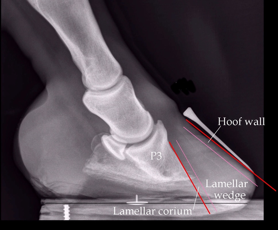
X-ray of a pony with long-term chronic laminitis showing a significant lamellar wedge
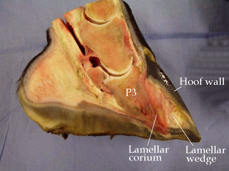
Photograph of a dissection of a horse euthanised because of laminitis, showing the lamellar wedge between the hoof wall and the lamellar corium (or dermal lamellae)
|
The external view of the foot in the x-ray, taken on the same day
|
Navicular
Horses with navicular are sometimes diagnosed as having laminitis, and vice versa.
Foot care for both laminitis and navicular have a lot of similarities, such as having the hoof capsule correctly aligned to the pedal/coffin bone, ensuring the foot is well protected/cushioned and that the horse is comfortably walking heel first. Barefoot rehabilitation appears to be very successful, as shown by the many successful rehabilitations from navicular by Rockley Farm.
Long Toe Woes: Perspectives on Navicular with Dr. Robert Bowker - The Humble Hoof, May 2020
Digging for the Truth about Navicular Syndrome - Pete Ramey
Boots and Pads: A True Breakthrough in Healing - Pete Ramey
Is the Hoof Smart? Adaptability of the Equine Foot - Dr Debra Taylor
Physiological Trimming for a Healthy Equine Foot by Dr. Robert Bowker
Dr. Robert Bowker on Navicular Disease - Casie, The Naturally Healthy Horse, May 2014 updated Feb 2017
Navicular syndrome - www.horsevets.co.uk
Facebook support group Barefoot Method for Navicular (administered by Alicia Harlov of The Humble Hoof)
This video Flat, Flared Right Front Trim by Daisy Bicking describes a trim to bring the toe back and raise the palmar angle, that may be suitable for a horse with navicular or caudal foot pain.
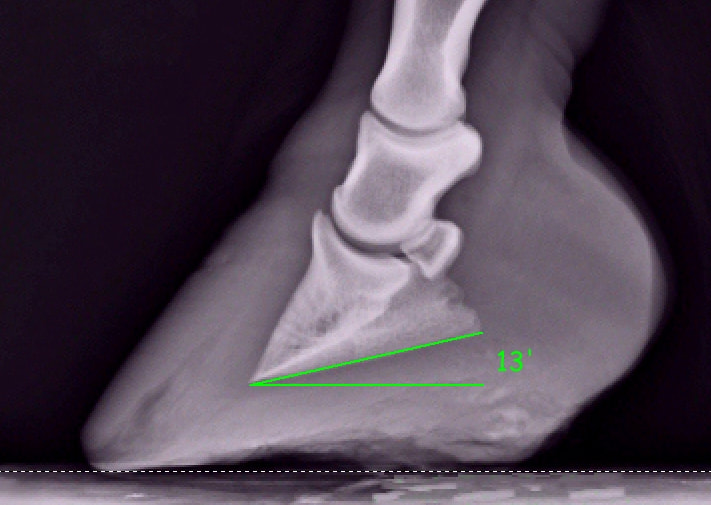
Palmar/plantar angle
The palmar or plantar angle (palmar relates to the front feet, plantar to the back feet) is the angle that the bottom (solar surface or distal border) of the pedal/coffin bone (P3) makes with the ground (as measured on a lateromedial radiograph). It describes the relationship of the pedal/coffin bone with the ground (or bearing surface).
There is lots of debate about what the ideal palmar/plantar angle should be. Suggestions include:
3 to 8 degrees - Debra Taylor 2011 in "Care and Rehabilitation of the Equine Foot" ("Most podiatrists agree that normal palmar/plantar angles range between 3-8 degrees").
2 to 10 degrees - Parks, Ovnicek and Sigafoos 2003 "The Foot and Shoeing, Diagnosis and Management of Lameness in the Horse".
3 to 5 degrees - Baxter, Shashak and Hill 2011 "Conformation and Movement" in Adams and Stashak's Lameness in Horses.
5 to 8 degrees - Daisy Bicking Hoof Guidelines
It seems to be reasonably widely accepted that a palmar/plantar angle between 3 and 8 degrees will be healthy/comfortable for most horses., and that the "correct" palmar/plantar angle will be that at which the hoof pastern axis or a line through the lower bones of the leg (P1, P2, P3) will be reasonably straight.
With navicular or caudal foot pain, the palmar/plantar angle is often too low, and may even become negative (i.e. the back of the bottom of P3 is lower than the front).
Following laminitis, the palmar/plantar angle often becomes too high, with heel height also too high.
Palmar Angles - The Equine Documentalist November 2020
Guidelines for Trimming the Equine Foot: A Review - Stephen O'Grady 2009
"Ideally, the palmar angle of the distal phalanx should be 3–5° greater than the dorsal angle of the distal phalanx, because this allows the distal phalanx to sink in a distopalmar direction during weight bearing and use the physiology in the palmar/plantar section of the foot. The palmar angle of the distal phalanx is generally dependent on the conformation of the palmar/plantar section of the foot."
Ramey, Pete
Pete Ramey's Articles - www.hoofrehab.com
"Most of the time the width of the true sole in front of the frog will be about 1/3 of the overall length of the frog" - p 344 Care and Rehabilitation of the Equine Foot - Pete Ramey.
Pete Ramey's Articles - www.hoofrehab.com
"Most of the time the width of the true sole in front of the frog will be about 1/3 of the overall length of the frog" - p 344 Care and Rehabilitation of the Equine Foot - Pete Ramey.
Rings and ridges
Hoof rings or growth rings are seen as minor lines in healthy hooves and are thought to be due to variations in nutrient content of the diet, particularly grass/forage. Slight colour variations are seen, but the hoof wall remains reasonably smooth with no significant change in texture.
Hoof ridges or fever rings can be seen and also felt as a distinct bump or ledge on the hoof wall, and are through to be suggestive of a systemic health problem, such as fever, laminitis, infection or toxicity. Often by the time the ridge is seen, the problem has been resolved.
Rings and Ridges: What Horse Hooves Reveal - KER - Feb 2016
Hoof Rings in Horses: What Do They Mean? - KER - Nov 2013
Shoes
Do shoes have a place in laminitis rehabilitation?
Possibly. Every horse, every situation is different, and what works for one horse may not work for another, and what works for a horse one month may not be the best option the next month. The most important part of foot rehabilitation following laminitis is trimming the hoof capsule to realign it with P3, thereby restoring P3 to its correct anatomical position, and keeping the trim correct.
"Anyone that puts a horse into a device without doing a correct trim is setting up the horse for failure in the recovery process." - EC & IR Group & Dr Eleanor Kellon - www.ecirhorse.com
Hoof boots and pads have developed significantly in the last few years and can usually be adapted to fit most rehabilitation scenarios.
Composite/plastic shoes have also developed significantly in recent years and many options are now available that give some sole protection/support. Composite shoes may be particularly useful a little time after the initial laminitis, when the foot is fully realigned and some laminar connections are re-established, so that the walls can take more loading. Also for shoes to be used, horses will need to be comfortable enough to stand on 3 feet, while the shoe is fitted.
EasyCare have developed several shoes suitable for therapy that can be used with sole support e.g. dental impression material and that can be glued, casted on nailed on, e.g.
EasyShoe Flex Light
Shoe A Horse With Super Glue - Garrett Ford April 2021 EasyCare Inc
Epona shoes have been used successfully in several complicated laminitis rehabilitations:
www.eponashoe.com
Hoof Help - UK distributor of Epona shoes
Hagen J, Hüppler M, Geiger SM, Mäder D, Häfner FS
Modifying the Height of Horseshoes: Effects of Wedge Shoes, Studs, and Rocker Shoes on the Phalangeal Alignment, Pressure Distribution, and Hoof-Ground Contact During Motion
Journal of Equine Veterinary Science , Volume 53 , 8 - 18 June 2017
"Pressure distribution showed wedges and studs to cause an increased pressure load on both the toe and the heels on a firm surface. Rocker shoes led to pressure peaks at the inner section of the toe, and high pressure was exerted on the quarters. In conclusion, all modified horseshoes showed unintended side effects and their influence on biomechanical parameters varied depending on the ground surface."
Heart bar shoes
Heart bar shoes - Nic Barker, Rockley Farm - May 2016
Heart bar shoes may remove the frog from action and lead to atrophy.
Reverse (Napoleon) shoes
Popular in Europe, the reverse shoe is a normal shoe fitted backwards, i.e. with the toe of the shoe under the heel of the horse, the idea being to bring breakover back and to remove forces on the laminae at the toe. However, if a realigning trim is not correctly carried out before the shoe is fitted, the shoe is unlikely to alter forces on the laminae to any great extent, and does not support P3 (see Care and Rehabilitation of the Equine Foot p244).
The effect of a reverse shoe and polystyrene padding on the biomechanics of the front hoof of the horse
Henning J Mostert - MSc dissertation 2009
|
Ski tip
Remodeling of the bone at the tip of the pedal/coffin bone (P3) is often referred to as a "ski tip". A ski tip is commonly seen following laminitis and may be a consequence of trauma, excess pressure on the rim of P3, decreased blood perfusion. |
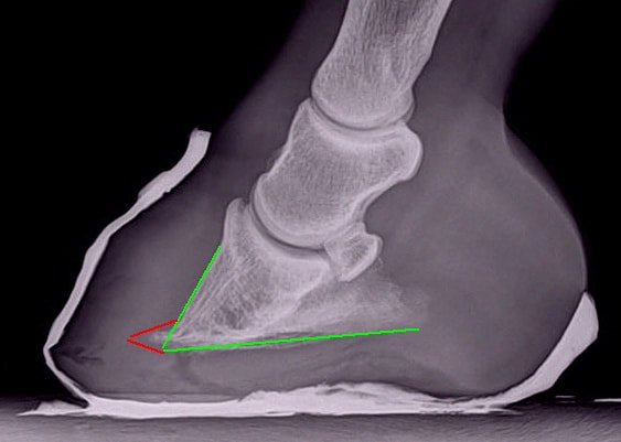
A long-term chronic laminitic foot. Green shows the likely correct outline of the pedal/coffin bone. Red shows the ski tip (bone remodeling).
|
Sole
Pete Ramey says that "the sole of the horse is the most important consideration in trimming" (Care and Rehabilitation of the Equine Foot p 284).
The sole is the barrier between the corium of P3 (the pedal/coffin bone) with its nerve and blood supply, and the ground, which may be hard or stony.
Managing thin soles - Linda Cowles www.healthyhoof.com
Sole ... "False" sole, shedding sole, retained sole. When to trim? When to leave it alone? - Linda Cowles www.healthyhoof.com
Uniform Sole Thickness (Researcher Explains How to Define a Heel) by M.T. Savoldi & G.F. Rosenberg - www.americanfarriers.org Jan/Feb 2003
Sole callous
Breakover- Pete Ramey
Soaking the feet may increase the moisture content of the sole:
Hampson BA, de Laat MA, Mills PC, Pollitt CC
Effect of environmental conditions on degree of hoof wall hydration in horses
Am J Vet Res. 2012 Mar;73(3):435-8. doi: 10.2460/ajvr.73.3.435
"Environmental conditions do not appear to affect moisture content in the hoof wall horn. Soaking horses' feet regularly in water would be unlikely to change the degree of hydration in the hoof wall horn but may further hydrate the sole."
Sole - necrosis and penetration
The sole grows from the solar corium (tissue containing blood vessels and nerves) on the solar surface (bottom) of P3 - see Horse Hoof Anatomy - www.all-natural-horse-care.com for a hoof dissection showing the solar corium.
The circumflex artery supplies blood to the sole, and lies close to the sharp solar edge of P3 - blood from the dorsal surface of P3 curls under the sharp edge of the bone to join the circumflex artery - see Colour Atlas of the Horse's Foot by Christopher C Pollitt, pages 19 - 24.
The solar corium with its blood and nerve supply lies between the hard pedal/coffin/P3 bone and the epidermal sole (and the ground), and is vulnerable to damage from compression. Compression from the descent and/or rotation of P3 (or any excess/constant pressure on the sole) can cause the sharp edge of the bone to reduce or cut off the blood supply to the solar corium, resulting in severe lameness and in some cases leading to necrosis of the sole (necrosis is defined as irreversible cell or tissue death from external factors - lack of blood supply can cause necrosis).
Source: Vet Clin North Am Equine Pract. 2010 Apr;26(1):29-49
The anatomy and physiology of the suspensory apparatus of the distal phalanx
Pollitt CC
For more websites showing the anatomy of the horse's foot including blood supply and sole corium, see Anatomy.
After The Crash - Lessons From Chronic Laminitis
Professor Christopher C Pollitt - available on www.safergrass.org
Slight rotation/descent of P3 can cause the sole to change from concave to flat. Blood vessels in the solar corium can be crushed by the sharp bottom edge of P3 and cause haemorrhage (NB use of medications that affect blood clotting, e.g. aspirin, can make haemorrhage more likely to occur), which may eventually appear as a crescent-shaped bruise as the sole grows down to ground level. After several weeks the tip of P3 can remodel and appear "ski-tipped" in lateromedial x-rays.
More significant rotation/descent of P3 can cause a convex bulge in the sole ("dropped sole"). The tip of P3 can crush the corium of the sole causing necrosis of the sole, and in severe cases P3 can prolapse/penetrate through the sole, often causing infection - abscess formation and infection of the bone (osteomyelitis). The tip of P3 can start to disappear (osteolysis). It is suggested that the hoof may grow inward towards the tip of P3, contributing to further rotation. The soft tissue and blood vessel compression and destruction of the bone is likely to cause significant pain and lameness, and can lead to incurable damage.
In time new yellow sole should replace the red necrotic solar corium under the tip of P3, and the reappearance of thick, concave sole is encouraging.
|
A correct realigning trim (above), protection and support of the foot and removal of the factors causing laminitis should generally prevent significant rotation (left) turning into P3 penetration of the sole (below).
|
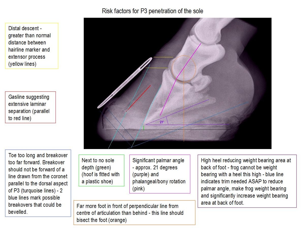
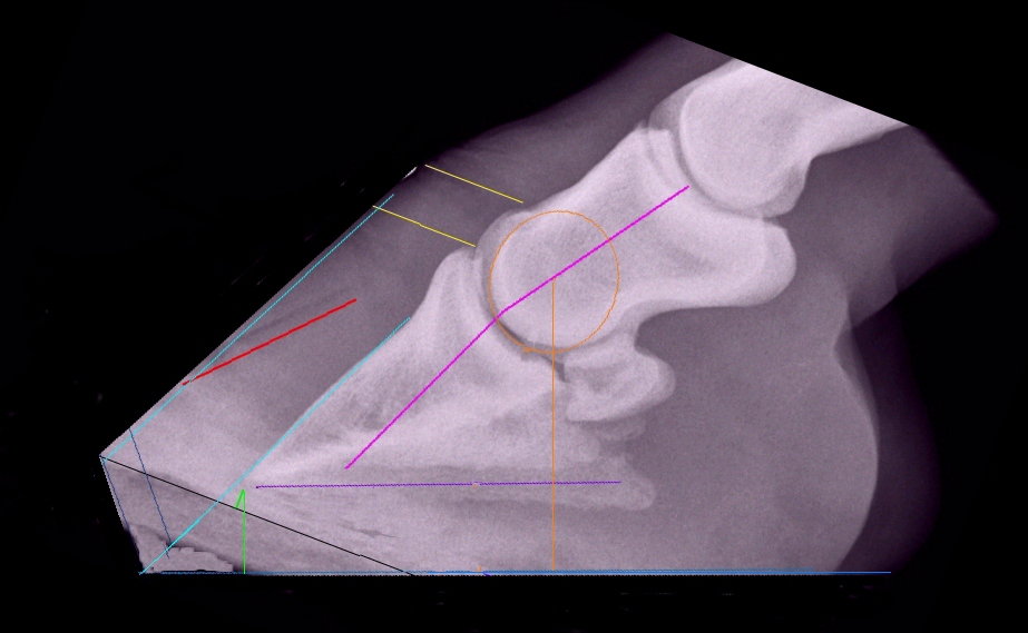
Trimming the foot with P3 penetration of the sole
The most important principles when trimming the foot with P3 penetration of the sole are:
The x-rays above show how trimming to lower the heels can immediately change a foot from one with significant pressure on the tip of P3 with subsequent compression of the sole and solar corium and the majority of weight being born on detached walls and at the front of the foot (above left) to one where the pressure on P3 is distributed widely and towards the back of the foot, and the majority of weight is being born at the back of the foot and shared by the frog, heels and bars (above right).
Radiographs (x-rays) should be taken immediately any signs of changes in the sole are seen (lateromedial x-rays should always be taken following laminitis).
The foot should be kept clean and disinfected, and probably covered/bandaged.
Auburn University Cases - www.hoofrehab.com - 2 cases presented with perforations on the sole at the tip of P3.
The Bars - Pete Ramey - www.hoofrehab.com - see photos dated 09/23/2006 and 11/3/2006 - before and after trimming of a hoof where P3 was lower than any part of the hoof wall and covered with only 3/16th inch of sole.
The most important principles when trimming the foot with P3 penetration of the sole are:
- to provide a good base of support at the heels, with the frog bearing weight;
- to reduce weight bearing on the detached wall;
- to relieve pressure on the exposed area.
The x-rays above show how trimming to lower the heels can immediately change a foot from one with significant pressure on the tip of P3 with subsequent compression of the sole and solar corium and the majority of weight being born on detached walls and at the front of the foot (above left) to one where the pressure on P3 is distributed widely and towards the back of the foot, and the majority of weight is being born at the back of the foot and shared by the frog, heels and bars (above right).
Radiographs (x-rays) should be taken immediately any signs of changes in the sole are seen (lateromedial x-rays should always be taken following laminitis).
The foot should be kept clean and disinfected, and probably covered/bandaged.
Auburn University Cases - www.hoofrehab.com - 2 cases presented with perforations on the sole at the tip of P3.
The Bars - Pete Ramey - www.hoofrehab.com - see photos dated 09/23/2006 and 11/3/2006 - before and after trimming of a hoof where P3 was lower than any part of the hoof wall and covered with only 3/16th inch of sole.
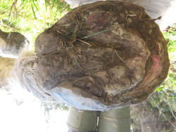
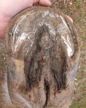
Some examples of sole penetration by P3 and successful rehabilitations (NB links are given purely to illustrate how sole penetration by P3 can look at various stages of rehabilitation):
Whisky - www.barehoofcare.com
P3 penetration in all 4 feet. His heels were lowered so that his frogs were weight bearing and his walls totally relieved from weight bearing. With frequent trims and good wound management, he was sound enough to go home after 4 months.
Glynn - www.naturalhorseworld.com - P3 penetration in all 4 feet.
Sole penetration - www.naturalhorsetrim.com
Bob - www.naturalhorsetrim.com
Druid - www.ironfreehoof.com - note the gasline in Druid's x-ray.
Rehabilitations - www.hoofrehab.com (Pete Ramey) - several rehabilitations following P3 penetration of the sole
Retracted sole
A retracted sole is when the sole appears to retract or be sucked up into the arch of the pedal bone (P3). The sole is often concave but very thin near the tip of the frog, rising up to a ridge/callous near the toe. Horses will often be sore and sensitive to sole pressure.
Retracted Soles: A Broader Perspective - Daisy Bicking Dec 2016
Concave but thin soles - being labelled "retracted soles" by some - may need careful trimming to avoid the feet becoming sore. Daisy Bicking suggests leaving more vertical height in the foot, not rolling the toe back too far into the callus, and protecting the feet with boots/soft pads.
Retracted Soles - Scoot Boots
My Horse Has Retracted Soles? Daisy Bicking Dec 2012 EasyCare Inc
Recognizing And Treating Retracted Soles - Esco Buff, Oct/Nov 2012 American Farriers Journal
Taylor, Debra
Taylor D, Sperandeo A, Schumacher J, Passler T, Wooldridge A, Bell R, Cooner A, Guidry L, Matz-Creel H, Ramey I, Ramey P
Clinical Outcome of 14 Obese, Laminitic Horses Managed with the Same Rehabilitation Protocol
Journal of Equine Veterinary Science Volume 34, Issue 4, Pages 556–564, April 2014 (online 05 Feb 2014)
Is the Hoof Smart? Adaptability of the Equine Foot - Dr Debra Taylor (video)
Taylor, Debra R
Laminitis Rehabilitation: If The Corium Is Happy, There Is Hope
Western Veterinary Conference 2013
Laminitis Rehab: 'If the Corium is Happy, There's Hope' - Erica Larson, www.thehorse.com Apr 2013
Taylor, Debra R
Every Hoof has a Story: An In-Depth Look at the Physical Examination of the Equine Hoof
Western Veterinary Conference 2013
Physical Exam of the Horse Hoof - Erica Larson, www.thehorse.com July 2013
CHAQUE PIED a une histoire : un regard en profondeur sur les examens physiques du sabot du cheval
Taylor, Debra R
Focus on the Ground Plane (Palmar/Plantar Angle)
Western Veterinary Conference 2013
Take The Plantar Angle Into Consideration - Jen Bradley, American Farriers Journal Sept 2014
Rouben CM, Taylor DR, Degraves FJ, Schumacher J, Guidry LN
Evaluation of the shape and depth of the collateral groove of the foot as a method to predict the position of the distal phalanx within the hoof capsule
Phi Zeta Research Day Forum 2012 p 37
Thrush
Thrush is very common in horses with laminitis, and is a common cause of lameness including what is often thought to be navicular disease. Feet with a deep central sulcus, often a result of heel contraction, are particularly prone to thrush.
Thrush is very common in horses with laminitis, and is a common cause of lameness including what is often thought to be navicular disease. Feet with a deep central sulcus, often a result of heel contraction, are particularly prone to thrush.
Underrun contracted heels have caused a deep central sulcus with a deep-seated thrush infection.
|
A healthy frog, shallow central sulcus, no signs of thrush.
|
Thrush is an infection (bacterial/fungal) of the frog and its sulci. There is often, but not always, a distinctive "cheesy" smell, black or grey discharge and spongy frog material, but any frog sensitivity may indicate thrush - the frog is often very sensitive to slight pressure, meaning that the horse will not land heel first.
Thrush can occur in wet or dry environments, wet alone is unlikely to cause thrush, although bacterial/fungal infections may be able to invade a soft frog more easily. The horse's environment should be kept clear of dung and urine, and a dry standing area provided - pea gravel is perfect for this, and provides stimulation for the frog.
Always consider the diet when a horse has thrush. Poor diet - nutrient deficiency, mineral imbalance, excess sugar/starch - may affect the skin and hooves. Illness and stress may also have an impact on skin and hooves. Owners often report thrush clearing up when zinc and copper deficiencies are addressed - nearly all forage diets are deficient in copper and zinc unless minerals are supplemented.
Frog stimulation and maximisation of circulation through heel first landing will reduce the incidence of thrush - the foot should always be trimmed to (eventually) maximise heel first landing, and the use of boots with thick pads can often make the horse comfortable enough to walk heel first.
Treatment of thrush
Many treatments sold for thrush will kill living tissue as well as the pathogens causing the thrush, often making the problem worse. If you wouldn't use the product on a sensitive area of your skin, do not use it on your horse's frogs. Treatments that shouldn't be used include formaldehyde, hydrogen peroxide, bleach and strong iodine - see the last paragraph of Linda Cowles' article: Thrush Treatment Summary.
Any treatment must get to the bottom of the sulcus.
Recommended treatments:
Soaking with CleanTrax - alongside correct trimming, this is often all that's needed in our experience.
Other treatments that have been suggested:
Soaking with White Lightning.
Mastitis medication, e.g. Albadry Plus (penicillin and novobiocin sodium).
Pete's Goo - 50% triple antibiotic ointment and 50% athletes foot cream (1% Clotrimazole) (NB antiobiotic creams cannot be sold over the counter in many European countries), or 50/50 mixture of 40% zinc oxide cream/Athletes Foot Cream (1% Clotrimazole). (Note: a mix of 40% zinc oxide cream with a small amount of copper sulphate powder is also suggested; however having seen a docile horse become violent when copper sulphate was applied to deep thrush, The Laminitis Site does not recommend the use of copper sulphate for thrush).
No Thrush/NT-Dry - use with care as this product is extremely drying.
Any treatment must be coupled with correct trimming, frog stimulation, diet, environment and hygiene.
Thrush Treatment - Pete Ramey
Frogs - www.wildheartshoofcare.com
Thrush - www.healthyhoof.com - Linda Cowles
Thrush Treatment Summary - www.healthyhoof.com - Linda Cowles
Frog Management - Pete Ramey - www.hoofrehab.com
Why does my horse land toe first? - Pete Ramey - Horseback Magazine July 2013
If I only had 4 frogs - Dawn Willoughby - Easycare Inc.
Learn to recognise a healthy frog: International Healthy Frogs Make Healthy Feet - www.healthyhoof.com - Linda Cowles
Toe
The wall at the toe should be parallel to the dorsal surface of the pedal/coffin bone.
Toes - www.wildheartshoofcare.com
Finding the true toe following laminitis
If there is laminar wedge, the position of the true toe (or where the toe should be) may not be obvious.
Ideally x-rays should be taken and used to guide the trim. The true toe will run parallel to the dorsal surface of P3.
When this is not possible, suggestions for finding the approximate position of the true toe include:
1. The 20-30 mm of hoof wall growth beneath the coronary band at the toe will often be "correct", i.e. parallel to the dorsal surface of P3, and this can be projected to the ground to show the true toe line.
2. Find the true frog apex. Approximately 2/3 of the length of the foot should be behind the true frog apex, and 1/3 in front.
3. Find the true frog apex. "Most of the time the width of the true sole in front of the frog will be about 1/3 of the overall length of the frog" - Pete Ramey, Care and Rehabilitation of the Equine Foot, p 344.
4. The hoof pastern axis may guide the position of the true toe - the toe should be parallel to the hoof pastern axis.
Toe Rocker
A toe rocker (bevel) reduces separating forces and encourages a tight connection between the pedal/coffin bone and the hoof wall.
Toe Rocker - all-natural-horse-care.com
Forward Foot Syndrome - Walter Friedrich
Walls & wall flare
Minimizing Hoof Capsule Distortion: How Minor Matters? - Daisy Bicking - EasyCare blog 2012
Hampson BA, de Laat MA, Mills PC, Pollitt CC
Effect of environmental conditions on degree of hoof wall hydration in horses
Am J Vet Res. 2012 Mar;73(3):435-8. doi: 10.2460/ajvr.73.3.435
"Environmental conditions do not appear to affect moisture content in the hoof wall horn. Soaking horses' feet regularly in water would be unlikely to change the degree of hydration in the hoof wall horn but may further hydrate the sole."
|
White Line The white line isn't white! When seen on the sole of the foot, the white line is a yellow/light tan colour. What looks like a white line is the stratum medium/inner hoof wall (sometimes called the water line). The white line is formed by the epidermal laminae (growing down past P3 with the hoof wall) and keratinised epidermal cells generated by the terminal papillae of the dermal laminae. See page 7 of Update on the anatomy of the inner hoof wall by Chris Pollitt for a diagram showing the terminal papillae of the dermal laminae (lamellae). 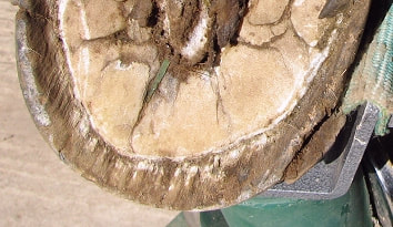
Stretched white line following laminitis
|
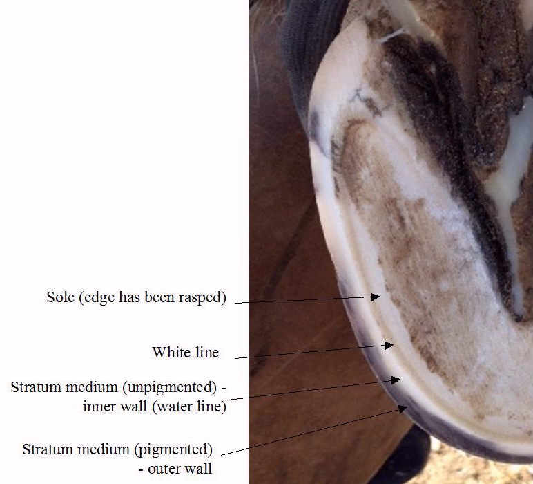
A tight healthy white line
|
|
Bruising/blood in the white line Bruising is caused by a trauma such as a blow or impact that ruptures underlying blood vessels, causing blood to escape into the surrounding tissues. The trauma that caused the bruising may have happened weeks or even months before the bruising is seen at the outer surface of the hoof. Bruising in the white line can be caused by: 1. Laminitis - red or pink areas of bruising, or pockets of dried blood or serum (yellow) can be seen in the white line following laminitis. If the white line is stretched, this may be more suggestive of the bruising being as a result of laminitis. 2. Anything that results in overloading the toe/toe first landing e.g. heel pain, high heels, upright feet, long toe/low heel conformation. If the white line is tight, this may be more suggestive of the bruising not being associated with laminitis. 3. Long toes that cause increased separating forces on the laminae. There may or may not be stretching of the white line. Hard, uneven and/or stony ground are likely to exacerbate bruising of the feet. More information: See The Essential Hoof Book - Susan Kauffmann, Christina Cline |
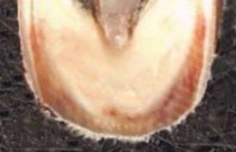
Blood and serum in the stretched white line of a laminitic foot
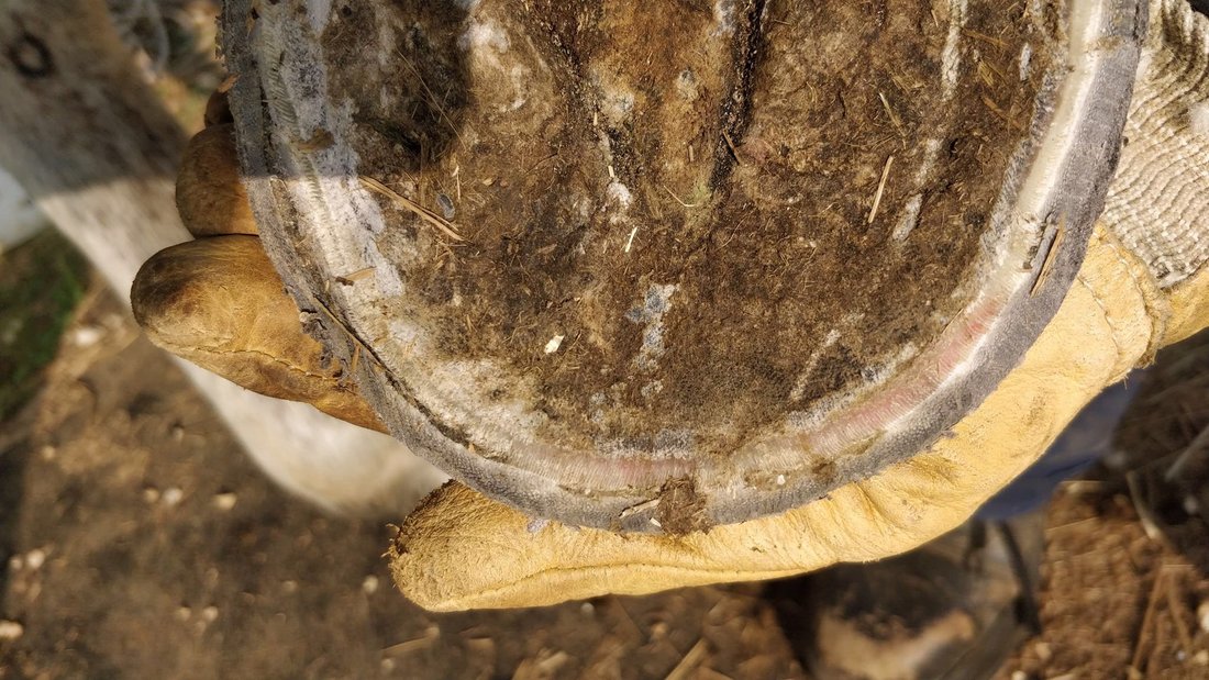
Bruising in a reasonably tight white line, possibly due to toe first landing and shallow sole depth/flat soles during an extended drought period with hard ground
|
Hoof Bruises - Paige Poss - www.ironfreehoof.com
Winter Leaving Its Mark - Christoph Schork - Easycare blog Feb 2011
This article has a photo of blood in the white line at the quarters, and the author says that he often sees blood in the white line in the quarters in early spring, and suggests that it may be due to the winter's icy frozen ground and subsequent concussion, perhaps due to an imbalance, but that the horses showing this blood in the white line are never lame.
White line / hoof wall separation
Separation of the hoof wall from the sole/widening of the white line is commonly seen following laminitis (including sub-clinical laminitis)* or when mechanical forces act on an imbalanced or overgrown hoof (e.g. long toes, overgrown walls, underrun heels), and can happen at the toe, quarters and/or the heels. Environmental conditions may also contribute. If the separation is invaded by bacteria/fungi, this can lead to white line disease. Where there is hoof wall/white line separation on the bottom of the foot, there may also be flared wall on the outside of the foot.
*Endocrinopathic laminitis often comes on insidiously and signs of lameness may not be seen, but sugar in the diet of a horse with insulin dysregulation may lead to sub-clinical laminitis and white line separation. If white line separation is seen after a period when a horse has had access to grass that could contain a lot of sugar, it would be sensible to consider endocrinopathic laminitis as a cause.
To improve white line separation, keep the feet well trimmed with frequent small trims if necessary, and (for bare feet) put a steep bevel on the outer wall where there is separation, starting 3 to 5 mm outside the true edge of the sole, i.e. just to the outside of or at the outer edge of the true white line. Protect the feet if necessary with boots and thick pads, particularly if the bevel decreases wall height. This series of photos shows a trim to correct a stretched white line: Toe Rocker - www.all-natural-horse-care.com.
White Line Disease
White line disease (WLD) can affect the white line and the stratum medium (inner hoof wall).
WLD may be mistaken for an abscess. Using x-rays can help differentiate (a 65 degree DP radiograph may be necessary) - gas shadows adjacent to the blood vessels will indicate an abscess.
In Care and Rehabilitation of the Equine Foot (p 337) Pete Ramey suggests that pathogens (bacteria, fungi) rarely affect healthy feet; often a wall crack or white line separation following laminitis allows pathogens to get established. Soak the foot with a solution that kills bacteria and fungi without damaging live tissue (e.g. CleanTrax, White Lightning, 50:50 apple cider vinegar and water) until the problem has fully resolved, and address the cause (diet, trim). Opening the lesion can be effective but may risk damaging the corium.
The end of white line disease - Pete Ramey 2005
Provide a dry environment, low sugar/starch mineral balanced diet and correct trim - remove flare, bevel walls, ensure the back of the foot is healthy and the horse is landing heel first (keep heels just above live sole, allow frog to develop, use boots and pads if necessary), treat fungal/bacterial infections.
Key points to consider for white line disease:
Discuss with vet and hoofcare provider.
Have radiographs (x-rays) taken.
Ensure feet are fully aligned to P3 (the pedal/coffin bone) and breakover/outer wall bevel is acting to reduce mechanical separating forces on the hoof wall, white line and lamellae/laminae. Keep feet well trimmed at all times so that cracks and separations cannot become significant.
Soak feet with CleanTrax.
Ensure the diet is providing NRC recommended levels of zinc and copper, balanced between 3:1 and 5:1 zinc:copper, and sufficient quality protein.
O’Grady SE & Burns TD
White line disease: A review (1998–2018)
Equine Vet Educ Feb 2021, vol 33 pages 102-112. Published online Oct 2020. doi.org/10.1111/eve.13201
"Improving hoof conformation and correcting any hoof capsule distortion that may have contributed to the hoof wall separation is the initial step in treating WLD. In order to prevent small separations from progressing, the clinician should examine each foot carefully during routine trimming. Separations involving the inner hoof wall should be explored and debrided down to solid horn whenever possible and any cavity that is left after debridement should be removed. If there are solid hoof structures on either side of the separation, the separated area is removed by using the rasp at steep angle and trimming the outer wall down to a solid sole-wall junction."
A fresh look at white line disease - Stephen E. O'Grady - Equine Veterinary Education Vol 23 Oct 2011
Management of White Line Disease - Stephen E. O'Grady - AAEP 2006
White Line Disease in Horses - Christa Lesté-Lasserre - www.thehorse.com - Aug 2010
White Line Disease - Ric Redden Jan 2009
Radiographic comparison of foot with WLD and foot with gas pocket following laminitis
WLD and nutrition
Feeding diets low in copper and zinc has been associated with a high incidence of WLD:
Higami A
Occurrence of White Line Disease in Performance Horses Fed on Low-Zinc and Low-Copper Diets
J Equine Sci Vol. 10 No. 1 p. 1-5 1999
Case studies of WLD with rotation of P3
Can Vet J. 2003 February; 44(2): 145–146.
Unilateral white line disease and laminitis in a quarter horse mare
Ray A. Oke
White Line Disease Case Report
Kate Echeverria, DVM 2010(NCSU), Richard A. Mansmann, VMD, PhD, Kurt Vom Orde and Andrew Cope
November 18, 2009
White Line Disease and Laminitis in a horse - Michael Porter - April 2013
White Line Disease - Equine Clinical Medicine, Surgery and Reproduction (p 73) - Graham Munroe, Scott Weese 2011
Instructions for trimming the toe to relieve WLD:
Physiological trimming for a healthy equine foot
Robert Bowker
Widest part of the foot
The widest part of the foot - Gene Ovnicek
No more than 50% of the foot should be in front of the widest part (measuring from breakover to the rear most weight bearing structure, i.e. the frog buttress).
The widest part of the foot can be found:
- around 1" back from the true apex (on a medium - size 0-2 - foot);
- where the bars terminate next to the frog;
- by marking an arc on both sides of the foot and seeing where the widest part is/measuring to see where the foot is widest.
Looking at radiographs, Ovnicek found that the widest part of the foot was on average 1/4" from the CoA on medium sized feet, and found that the relationship of the CoA with the widest part of the foot depended on the palmar/plantar angle (he found that with a negative to 5 degree palmar angle the CoA was behind the WPOTF, once the palmar angle was 6+ degrees the CoA was in front of the WPOFT).