Chronic laminitis
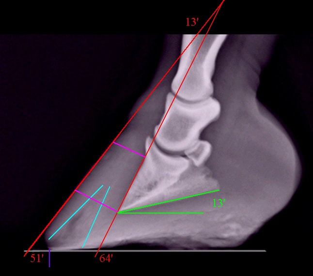
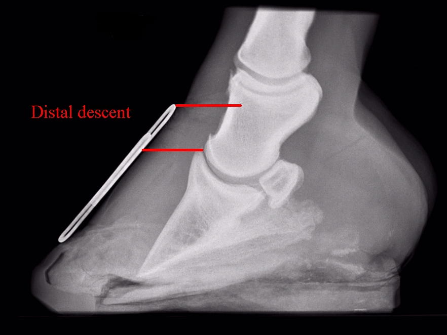
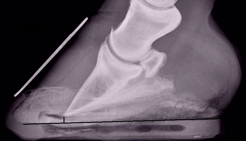
Chronic laminitis means that laminitis / laminopathy has taken place in the feet and the consequent damage has not been fully corrected, i.e. there has been some loss of correct alignment between the hoof capsule and the pedal/coffin bone. This might be dorsal rotation, palmar (plantar if on a hind foot) rotation, sinking (distal descent), abnormally thin soles, remodeling of P3 (particularly at the tip).....
In Equine Laminitis (Belknap, Wiley Blackwell 2017) [1], Philip Johnson gives this definition: "Chronic laminitis represents the situation in which disease of the hoof lamellar interface has resulted in abnormal hoof growth or deformity and may nor may not be associated with pain (lameness)."
It is not unusual for horses with laminitis due to insulin dysregulation (EMS / PPID) to have chronic laminitis without having shown signs of being lame - according to Ninja Karikoski [2], "naturally occurring endocrinopathic laminitis is a chronic process" and laminitic "lesions may have been developing for months or even years". It is very important that owners, vets and hoofcare professionals can recognise the signs of chronic laminitis, which, if seen, should instigate the taking of x-rays of all affected feet to guide realigning trimming, plus good support/protection of the feet.
Divergent hoof rings (wider at the heels than at the toe) and a stretched or separated white line (sometimes with signs of blood or serum) are generally accepted as signs of chronic laminitis, and x-rays should always be taken if these signs are seen.
Symptoms of chronic laminitis
|
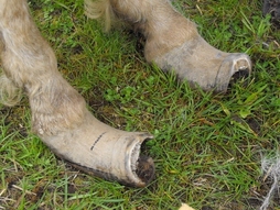
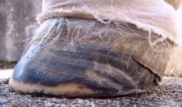
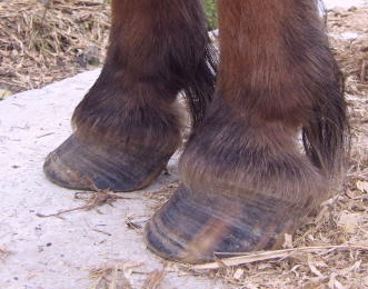
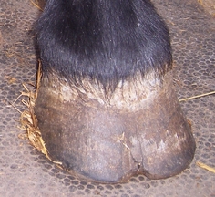
Abnormal hoof growth - this is extreme but does happen when feet are neglected. See Cedar's amazing rehab for a happy ending.
|
|
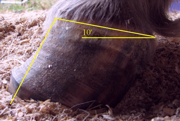
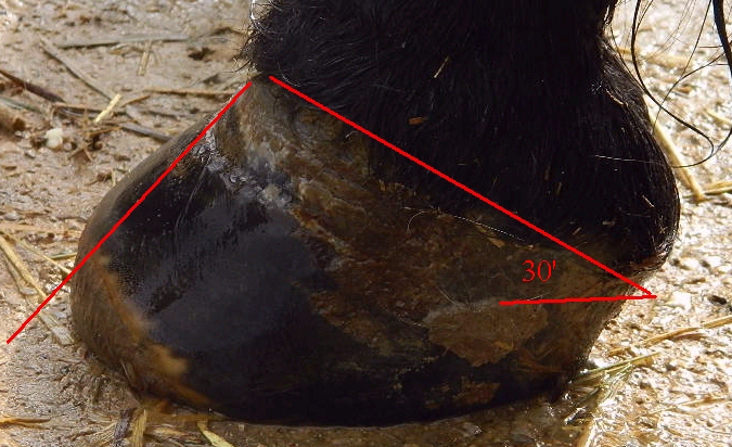
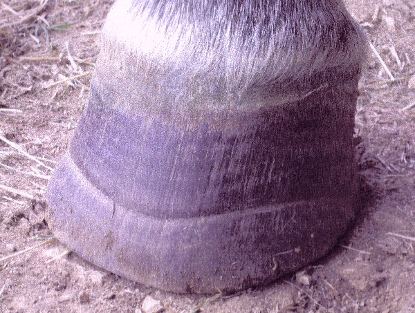
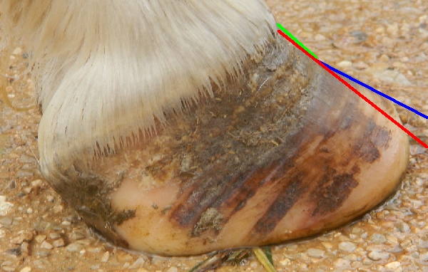
Heels grow quicker than toes, often causing a "boxy" appearance to the hoof with the hair line straighter than normal from toe to heel when viewed from the side (yellow line - left).
Compare to the well trimmed laminitic hoof with sloping hair line (red line - right). |
|
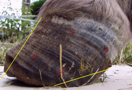
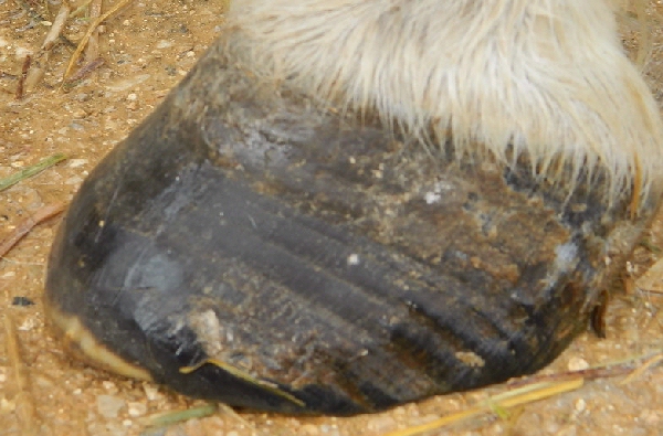
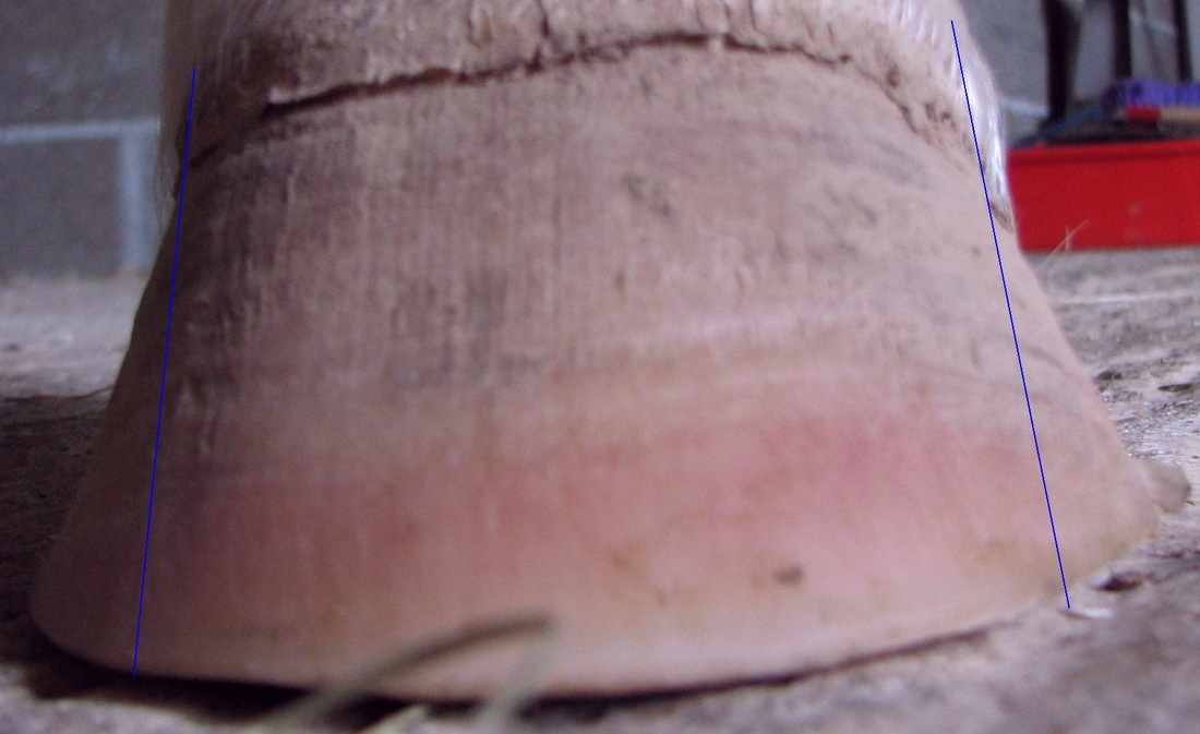
Divergent hoof rings - rings are wider or lower at the heels than at the toes.
Rings on the hoof wall from coronet to ground are an indicator of ongoing chronic/endocrinopathic laminitis. Horses with distal descent (sinking) often have a hoof ring the same distance from the coronet the whole way around the hoof. It takes a minimum of 3 months for new divergent hoof rings to be seen following laminitis (1). Horses can have growth rings as a result of changes in diet or environment rather than laminitis - these will not be divergent. |
|
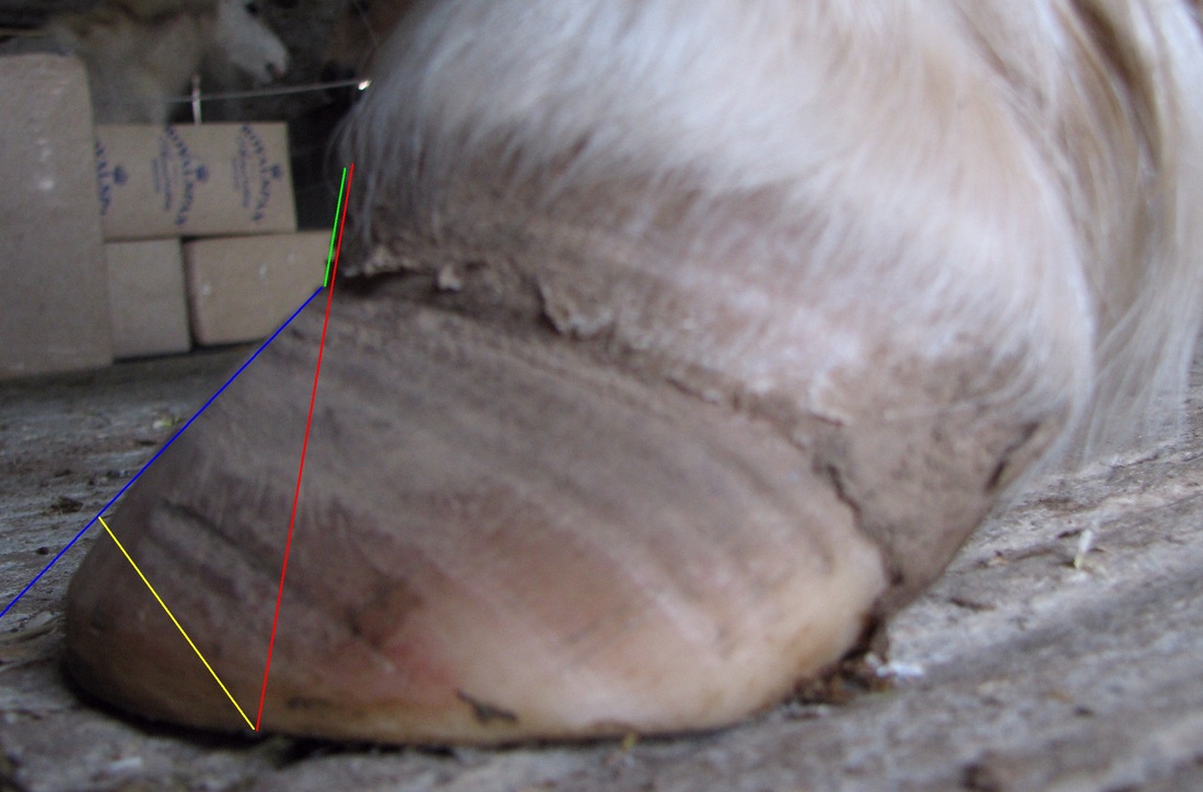
Change in hoof wall angle - tight new growth directly below the coronet (green), flared wall with loose laminar connections (blue). Trim required to encourage new tight growth (bevel - yellow, line of new growth - red). Bell shape or bulge in hoof wall. Bruising/redness in hoof walls. Flared walls. Wall cracks. |
|
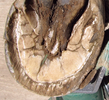
Deep black groove between wall and sole where tight white line should be.
Blood/haemorrhage in white line. |
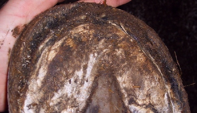
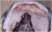
Stretched white line (stretched epidermal laminae).
|
Solid laminar wedge can fill the gap between separated wall and sole and can look just like sole, but is often a slightly different colour and often the outline of P3 can be seen.
|
|
|
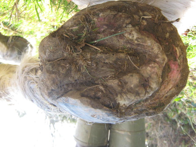
Bruising on the sole in front of the frog and below the tip/solar edge of P3.
Bulging/convex/dropped sole (photo p61). Depression just above the coronet (photo p61) - as you run your finger down the front of the pastern towards the hoof, your finger "gets stuck" in a dip just above the start of the hoof wall.
Swelling/deformation at the coronary band (photo p2) Penetration of sole by P3 - solar prolapse.
|
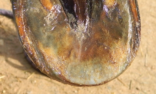
Thin sole (above)
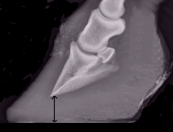
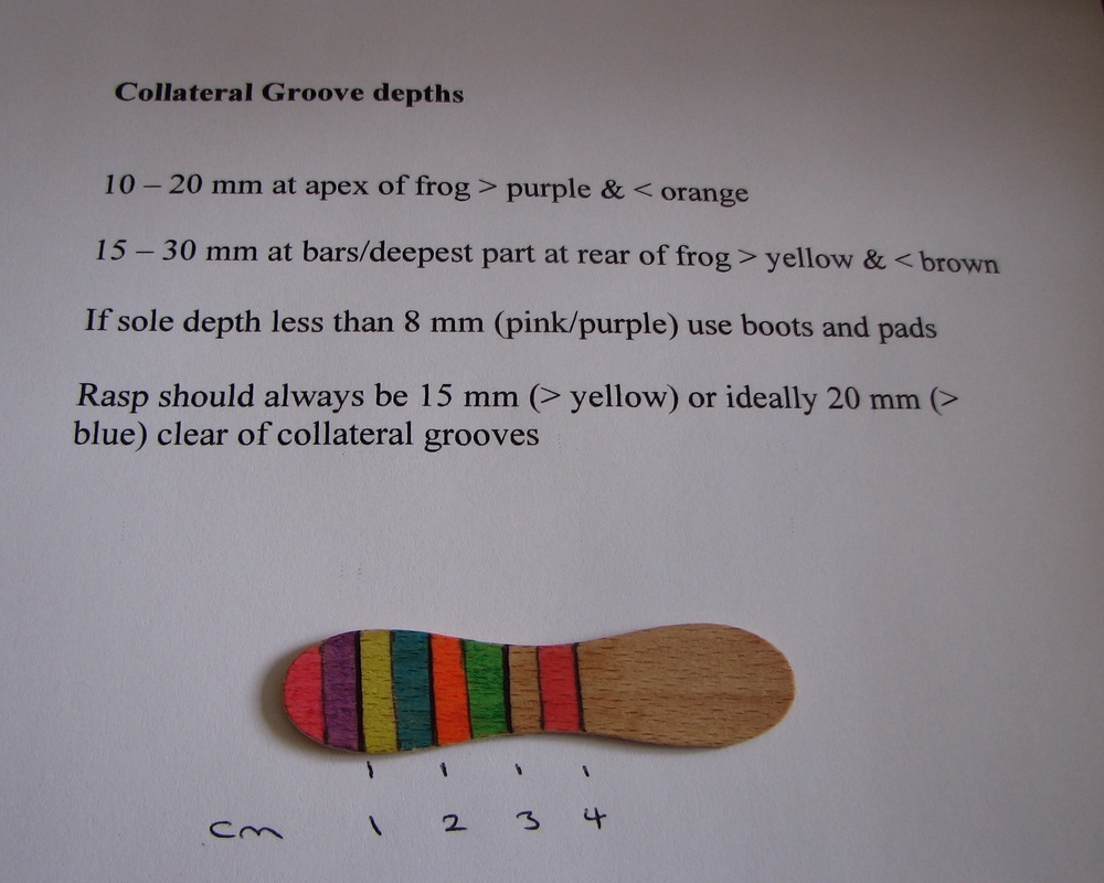
or thick sole (below) - thickened sole seems to be more common in ponies. Sole thickness can be estimated by measuring the collateral groove depth. |
|
Large difference in collateral groove depth between front and back of frog.
For most horses a good collateral groove depth at the apex of the frog (measured to the sole by the white line) will be 10-20 mm - less than 10mm is likely to indicate a thin sole; and 15-30 mm at the deepest part (usually near the bars). The greater the difference between the collateral groove measurement at the apex and the back of the frog, the greater the palmar angle and therefore P3 rotation. |
Further information
After the Crash - Lessons from Chronic Laminitis - Professor Christopher C Pollitt BVSc, PhD (www.safergrass.org)
References
[1] Equine Laminitis 2017 edited by James Belknap, published by Wiley Blackwell
[2] Ninja Karikoski Dissertation May 2016
The prevalence and histopathology of endocrinopathic laminitis in horses
