
Farriers and trimmers often worry about shortening the toe on a horse with dorsal rotation, thinking it could cause pain. These diagrams hopefully explain why it doesn't....
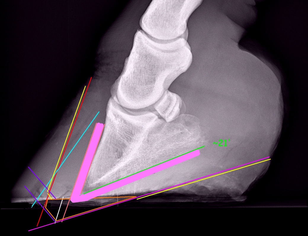
Above: The only nerves and blood vessels in the front of the foot are in the coriums that surround P3 - shown by the thick pink lines. The hoof to be trimmed is marked in yellow, the hoof to protect and grow is marked in orange. You can see that the pink area falls wholly within the orange area where we need to protect the foot, and nowhere near the yellow area where we want to trim the foot. The only exception to this is where the tip of P3 has some bone remodeling - there does appear to be a bit of remodeling on this x-ray - so allowing a slight margin for this is sensible, but even allowing for a decent margin around the tip of P3, the yellow trim lines aren't near the corium/tip of P3.
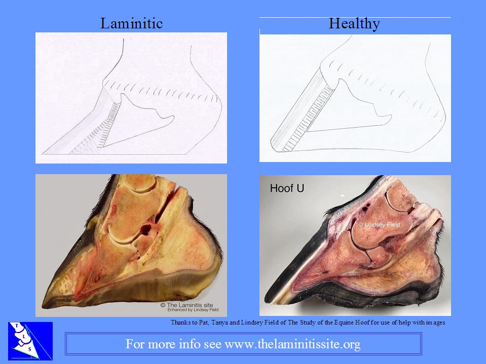
Above: This diagram shows a drawing and dissection of a laminitic foot (left) against a healthy foot (right).
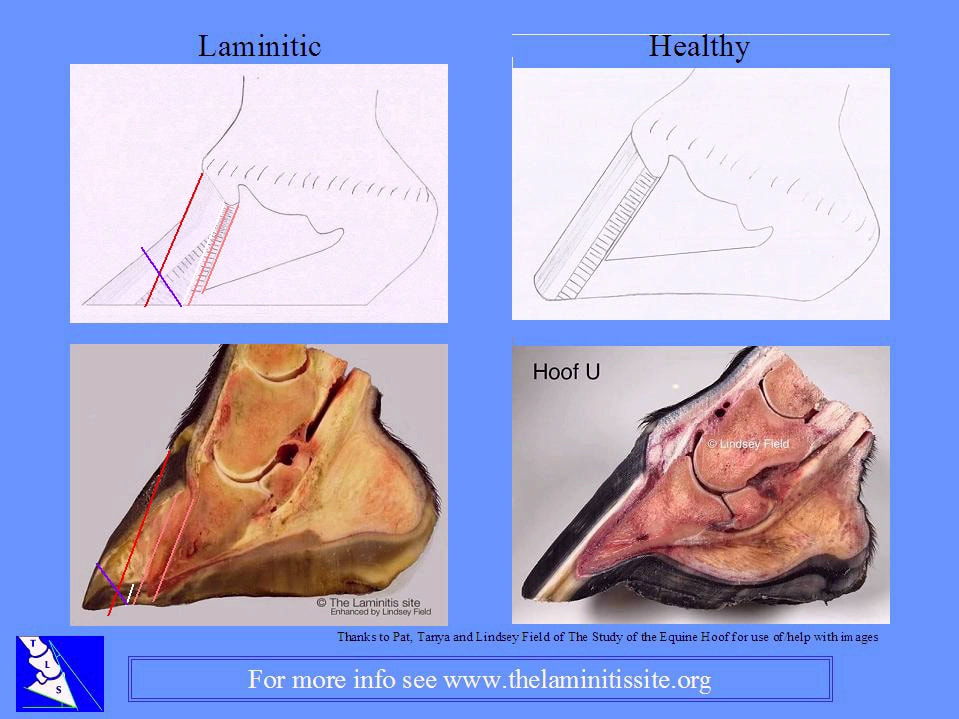
Above: The same coloured lines are marked on the diagram and photo of the laminitic foot to show how the trim doesn't go near the nerves and blood in the laminar corium:
Inner pink line = dorsal surface of P3.
Outer pink line = outer edge of dermal laminae and edge of sole at ground level.
White line = outer edge of white line (which is the outer edge of the epidermal laminae), being around 3 mm to the outside of the true edge of the sole.
Red = true toe - runs from the hairline at the toe to the ground, parallel to the dorsal surface of P3.
Purple = breakover bevel to reduce separating forces on the toe. Ideally this will start at or just to the outside of the edge of the true white line, so 3 to 5 mm outside of the true edge of the sole.
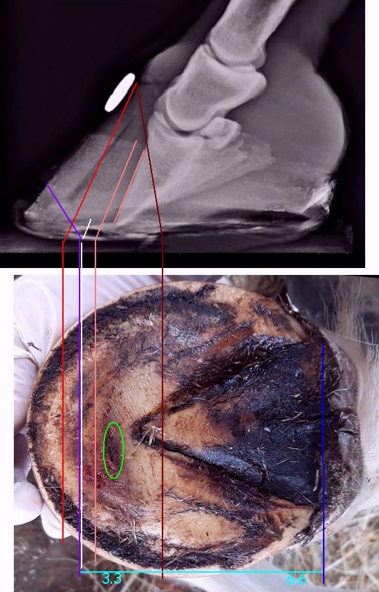
Above: Note we talk about the "true" white line and sole, as the true white line and sole can be covered over by sole/laminar wedge material, as on this foot with a solar penetration below, taken around 2.5 months after the sepsis-related laminitis event - so the foot has had time to change/grow abnormally. The x-ray and sole photo are lined up reasonably well (but not exactly). The position of the tip of the pedal bone can be seen outlined in green (this may be very slightly in front of the tip of P3 - it's the opening in the sole where the tip of P3 appeared a couple of weeks later (presumably because the foot was not correctly realigned and supported)).
Brown - the frog apex may have grown very slightly forwards - but also may not have done - the true frog apex should be checked on the foot. This provides a good guide, as approximately 2/3 of the total length of the foot (to the back of/widest part of the frog) should be behind the true apex, and approximately 1/3 of the total length of the foot (to breakover) should be in front of the true apex - in the photo there are 3.3 units from purple breakover to brown frog apex, and 6.6 units from brown frog apex to blue back of frog.
Red - the true toe runs from the hairline to the ground parallel with the dorsal surface of P3.
Inner pink - marks the dorsal surface of P3.
Outer pink - suggests the approximate outer edge of the dermal laminae and the true edge of the sole at ground level. As there is almost no sole depth, the edge of the sole is very close to the tip of P3. See how the edge of the sole looks to be much further forwards - what looks like sole in the front of the foot is actually laminar wedge.
White - suggests the approximate outer edge of the white line (which is the outer edge of the epidermal laminae), being around 3 mm to the outside of the true edge of the sole. Again, see how it looks as if the white line is just behind the toe on the photo, where you would expect it. What looks like the white line on the sole view is the end of the epidermal laminae, which have been pushed away from the dermal laminae and the gap between filled with laminar wedge - you can see this in the dissection photo above (and see also the diagram of the laminitic foot in the previous images). The true white line will always be just outside the true edge of the sole, growing down from the terminal papillae where the dermal laminae reach the bottom of P3.
Purple - suggests a breakover bevel to reduce separating forces on the toe. Ideally this will start at or just to the outside of the edge of the true white line, so 3 to 5 mm outside of the true edge of the sole. Yes, this is very close to the tip of P3 - but that's because there's no sole depth.
All of these diagrams suggest an accurate - and therefore quite scary and severe - trim, because of the lack of sole depth bringing the trim so close to the tip of P3. It's great for a model. In reality it always pays to err on the side of caution, but keep in mind that not doing enough can be (almost) as bad as doing too much.
In reality, based on the foot shown in the top x-ray (showing the corium in thick pink) you might aim to get the true toe (the red line) rasped in the first trim - as long as the laminar wedge was stable - then put a very small (but steep) breakover bevel behind the true toe, to reduce the separating forces on the toe. Then in around 1-2 weeks, assuming that the bottom of the foot had been rasped into 2 clear planes at the first trim and the horse was now able to weight the back of the foot and relieve the front of the foot and sole, you would expect there to be a reasonable increase in sole depth in the front of the foot, and you might then take the breakover bevel back to or just outside of the edge of the true white line. The angle of the bevel is important - even a bevel just behind the true toe line, if set at a steep angle*, should help to reduce separating forces on the toe (and therefore pain and the risk of further damage).
(* aim for a bevel of around 60 degrees in the initial stages of rehabilitation, reducing to around 45 degrees as the foot improves, then finally to 30-45 degrees on the recovered foot).
Further information
For members of the ECIR group: Backing up the Toe - Eleanor Kellon August 2017
Toe Rocker - www.all-natural-horse-care.com
Laminitis And The Laminar Wedge: Take It Or Leave It
Using the Rasp (using and applying the bevel)
For members of the ECIR group: Backing up the Toe - Eleanor Kellon August 2017
Toe Rocker - www.all-natural-horse-care.com
Laminitis And The Laminar Wedge: Take It Or Leave It
Using the Rasp (using and applying the bevel)
 RSS Feed